Vision Centre
Southport and Robina
Gold Coast Queensland Australia
Fluorescein Angiogram
The Yellow Dye Test
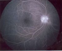
![]() A fluorescein angiogram is a yellow dye test used to detect changes and abnormalities in the retina. The dye is injected into your arm and once in the blood stream is able to be photographed in the vessels that are in the back of your eye. Fluorescein angiograms are used in conditions such as diabetic retinopathy and macula degeneration.
A fluorescein angiogram is a yellow dye test used to detect changes and abnormalities in the retina. The dye is injected into your arm and once in the blood stream is able to be photographed in the vessels that are in the back of your eye. Fluorescein angiograms are used in conditions such as diabetic retinopathy and macula degeneration.
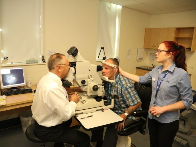
![]() The fluorescein angiogram (FA) is a diagnostic procedure using a special camera to take a series of photographs of the retina or back of the eye that can assist in the diagnosis of conditions affecting the back of the eye including diabetic retinopathy and macular degeneration as well as the suitability for different treatments for these diseases such as lucentis and laser. The FA exam can lead to earlier confirmation of damage or disease so doctors can provide treatment sooner. You will be given an information sheet prior to your appointment for the FA exam which will explain the need to wear a loose sleeve shirt that can be rolled up and not restrict the blood flow in your arm. You will be advised that you will need to be at Vision Centre for 1 to 2 hours on the day of the procedure.
The fluorescein angiogram (FA) is a diagnostic procedure using a special camera to take a series of photographs of the retina or back of the eye that can assist in the diagnosis of conditions affecting the back of the eye including diabetic retinopathy and macular degeneration as well as the suitability for different treatments for these diseases such as lucentis and laser. The FA exam can lead to earlier confirmation of damage or disease so doctors can provide treatment sooner. You will be given an information sheet prior to your appointment for the FA exam which will explain the need to wear a loose sleeve shirt that can be rolled up and not restrict the blood flow in your arm. You will be advised that you will need to be at Vision Centre for 1 to 2 hours on the day of the procedure.
![]() When you arrive for your fluorescein angiogram you will have dilating drops instilled. You will therefore be unable to drive after this test so it is requested that you have someone drive you home. After your pupils are dilated you will be seated at the FA camera and a special yellow vegetable dye (called fluorescein) will be injected into a vein in the arm. As the dye passes through the blood vessels at the back of the eye your doctor will take multiple photographs of the retina.
When you arrive for your fluorescein angiogram you will have dilating drops instilled. You will therefore be unable to drive after this test so it is requested that you have someone drive you home. After your pupils are dilated you will be seated at the FA camera and a special yellow vegetable dye (called fluorescein) will be injected into a vein in the arm. As the dye passes through the blood vessels at the back of the eye your doctor will take multiple photographs of the retina.
If you suspect or know you are allergic to vegetable dye please inform the doctor prior to your appointment.
The FA camera sends the photographs to the computer instantly so you will be able to view the results and discuss them with your Ophthalmologist. The fluorescein dye will then harmlessly filter out of your body through the kidneys over the next few hours.
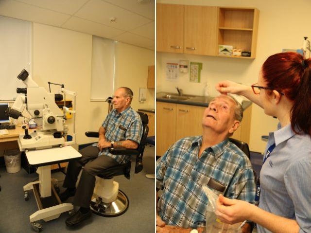
1. COMFORTABLE SEATING FOR DILATION OF YOUR EYE
2. PRELIMINARY EXAMINATION PRIOR TO PREPARING THE DYE
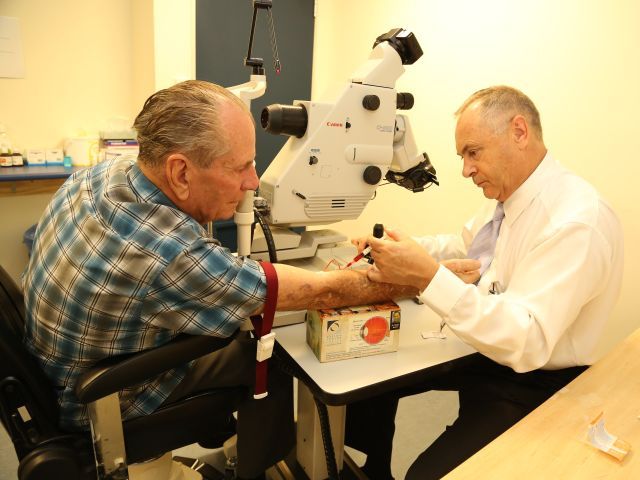
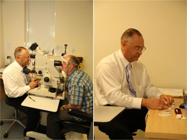
3. A QUICK PROCESS TO INTRODUCE THE DYE INTO YOUR BLOOD STREAM
4. MULTIPLE PHOTOGRAPHS ARE TAKEN OF THE RETINA IN YOUR EYE
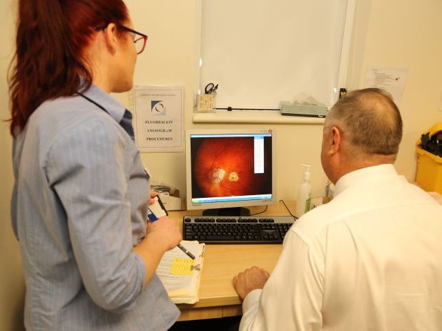
5. THE PHOTOGRAPHS TAKEN ARE CAREFULLY EXAMINED AND ASSESSED
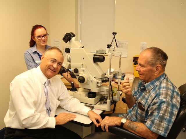
6. THE RESULTS ARE DISCUSSED IN A RELAXED BUT CARING ATMOSPHERE
![]() Top of Page
Top of Page![]()
Copyright © 2000+ All Rights Reserved